
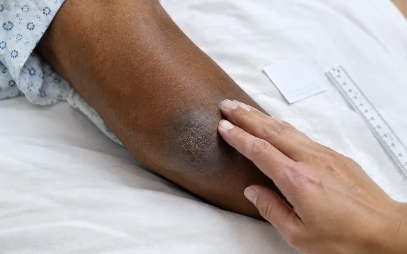
On lighter skin, an early bedsore is a red or pink patch over a bony spot that stays red after pressure comes off. On darker skin that redness often does not show. The same injury may look darker than the surrounding skin, or bluish or ashen, and feel warmer, cooler, firmer or softer. On deeper skin tones, touch and temperature tell you as much as color.
Most descriptions of bedsores start with redness, and redness is the sign most likely to be missed on brown and Black skin. For a family visiting someone who spends most of the day in a bed or chair, that gap matters. An early pressure injury usually heals once the pressure is relieved. A late one can mean months of wound care, infection or a hospital stay.
Why redness is the wrong test on darker skin
A pressure injury starts when body weight pins skin between a bone and a mattress or chair long enough to cut off its blood supply. The official staging definitions from the National Pressure Injury Advisory Panel (NPIAP) describe stage 1 as intact skin with redness that does not fade when pressed. They add that it can look different on darkly pigmented skin, and that changes in sensation, temperature or firmness can come before any visible change.
Melanin masks the pink flush that pale skin shows, and the quick press test, where healthy skin whitens briefly under a fingertip, is hard to read on deep skin tones. Cleveland Clinic notes the color change can be hard to see, and that the area may instead feel warmer, cooler, softer or firmer. The result is late discovery. A 2023 NPIAP review reports that pressure injuries on dark skin tones are more often identified at a later stage, and a scoping review in PubMed Central found routine visual checks unreliable for early detection in darker skin.
The scale is not small. In the CDC’s 2004 National Nursing Home Survey, about 159,000 residents, 11% of everyone living in US nursing homes, had a pressure ulcer, and stage 2 was the most common.
What each stage looks like
| Stage | What it looks like | What changes on darker skin |
| Stage 1 | Intact skin over a bony area with a red or pink patch that does not fade when pressed. | Redness may not show. Look for a patch darker than the surrounding skin, sometimes bluish or ashen, and check for warmth, coolness, firmness or tenderness. |
| Stage 2 | A shallow open sore with a moist pink or red base, or a fluid-filled blister, intact or broken. | The wound bed still looks pink or red. The skin around it may look darker than usual. |
| Stage 3 | A deeper crater through the full thickness of the skin, where fat may be visible. Yellowish dead tissue may cover part of it. | Looks broadly similar on all skin tones. |
| Stage 4 | A full-thickness wound with muscle, tendon or bone exposed or able to be felt. | Looks broadly similar on all skin tones. |
| Unstageable | The base is hidden under yellow, tan, gray or brown dead tissue or a dark, hard scab, so its depth cannot be judged. | Looks broadly similar on all skin tones. |
| Deep tissue pressure injury | A persistent deep red, maroon or purple area, or a dark blood-filled blister. Pain and temperature change often come first. | The discoloration can blend into natural pigment. Compare with the same spot on the other side of the body. |
.
Many guides describe a stage 1 bedsore on dark skin as purple. NPIAP draws the line differently: its stage 1 definition excludes purple or maroon discoloration, which it flags as a possible deep tissue pressure injury, damage where bone meets muscle that can open into a deep wound quickly. On darker skin the two are hard to separate by eye, so any new purple or maroon patch over a bony area warrants a nursing assessment the same day.
How to check skin during a visit
With the resident’s permission, and ideally with a nurse present, a skin check takes a few minutes:
- Start with the pressure points. The CDC lists the elbows, heels, hips, shoulders, back and back of the head as the usual sites. Add the tailbone and any skin under tubing or braces.
- Use strong, white light. Dim rooms and warm bulbs hide color shifts, and a hospital quality project on dark skin tones built its skin assessment around halogen lighting for that reason.
- Compare left with right. The same spot on the opposite hip or heel is the best reference for that person’s normal color and temperature.
- Feel as well as look. Use the back of the fingers to compare warmth, press gently for firmness or a spongy, boggy feel, and ask about pain, burning or itching.
- Moisturize dry, ashy skin before judging color. Wound nurses point out that an ashy film, calluses and dim light can all hide early damage on darker skin.
- Watch the clock. Johns Hopkins Medicine flags redness that is still present 30 minutes after pressure comes off, and advises people with dark skin to call a provider if a patch turns darker or feels warm.
Photograph and describe what you see
With consent from the resident or their representative, dated photos are the most useful record a family can keep. Color is also what a phone camera gets wrong most: auto white balance, filters and room light shift skin tone between pictures, making a wound look better or worse than it is. The following comes from color-matching practice rather than a clinical standard:
- Shoot in the same position and the same light every time, ideally daylight from a window, with filters and auto-enhance switched off.
- Place a plain white card beside the area, never on it, so color can be compared across photos, with a ruler beside it for size.
- Write down the person’s usual skin tone in plain words, depth and undertone, so a note like “the patch on her left hip is darker and cooler than her usual deep brown” means something to the next nurse or doctor. The skin tone chart and the list of skin color names give usable vocabulary.
- Keep the photos private and share them only with the care team, family decision-makers or an attorney.
Bedsore or moisture damage?
Not every sore area is a pressure injury. Urine, stool and sweat cause moisture-associated skin damage, a rash-like breakdown that tends to spread across the buttocks, groin or skin folds instead of sitting over a bone. NPIAP’s definitions say it should not be labeled stage 2. The two are connected, though: in the CDC survey, residents with recent incontinence had pressure ulcers more often than continent residents, 12% against 7%.
Prompt cleaning, gentle drying, a barrier cream and clean, dry bedding protect skin either way, the same logic behind skin health and environmental hygiene in any bedroom.
Why older skin breaks down faster
Skin thins with age, loses some of the fat that cushions bony points, and heals more slowly. Weight and movement shift the odds further: in the CDC survey, 20% of residents with recent weight loss had pressure ulcers against 10% of those without, and 16% of residents with high immobility had them against 5%. Nutrition, fluids and regular turning are part of prevention, and the everyday habits that support skin as it ages still apply to someone living in a care home.
What nursing homes are required to do
Federal rules for nursing homes certified by Medicare and Medicaid are specific. Under 42 CFR 483.25, a facility must provide care consistent with professional standards to prevent pressure ulcers, and a resident should not develop one unless their clinical condition shows it was unavoidable. A resident who has one must receive the treatment needed to promote healing, prevent infection and stop new ones forming.
The standard cuts both ways. Some pressure injuries are unavoidable in very ill residents, and one sore is not proof of neglect. A facility is still expected to document risk assessments, turning schedules, wound measurements and care plan changes, and families can ask to see them. In the 2004 CDC survey, only 35% of residents with stage 2 or worse ulcers had care from a specialized wound program.
If you think a bedsore was missed
Medical care comes first. Ask for a wound assessment by a nurse or physician, how the injury was staged and what the treatment and turning plan is, in writing.
Under the Older Americans Act, every state has a Long-Term Care Ombudsman Program, a free, confidential advocate that takes complaints about residents’ care. If a wound appeared or worsened without explanation, or treatment was slow, families weighing legal options can read nursing home bedsore lawsuit guidance that explains which records matter, how an injury timeline is built, and why state filing deadlines make early action important.
Questions families ask
What does a stage 1 bedsore look like on dark skin?
Usually a patch over a bony area that is darker than the surrounding skin, sometimes with a bluish or ashen cast. It may feel warmer or cooler, firmer or softer, and the person may report pain, burning or itching before anything is visible.
Is a purple patch on the skin a bedsore?
It can be. NPIAP treats persistent purple or maroon discoloration over a pressure point as a possible deep tissue pressure injury, which can open quickly, so it needs a nursing check the same day.
Is every bedsore a sign of neglect?
No. Federal rules allow that some are clinically unavoidable. The questions that matter are whether the facility assessed the risk, followed its care plan and treated the wound promptly.
Someone who depends on others to turn, wash and dress them also depends on others to notice their skin. A family that knows what early damage looks like on that person’s skin tone is one of the most reliable second checks they have.
How this was checked: stage descriptions follow the National Pressure Injury Advisory Panel’s official definitions; prevalence figures come from the CDC’s National Center for Health Statistics and date from its 2004 survey; the federal standard is summarized from 42 CFR 483.25. The photography guidance applies color-matching practice and is marked as practitioner judgement. This guide supports, and does not replace, assessment by a nurse or physician.

BeautyLiesTruth publishes hair and personal color guidance built on primary research. Color claims are tested by draping under controlled daylight, and every figure is traced to a published source rather than to another blog.